Long Bone Diagram Hyaline Cartilage : Structure Of A Typical Long Bone - Hyaline cartilage transmits sound waves fairly uniformly.
byAdmin-
0
Long Bone Diagram Hyaline Cartilage : Structure Of A Typical Long Bone - Hyaline cartilage transmits sound waves fairly uniformly.. On the diaphyseal side, cartilage is ossified, and the diaphysis grows in length. Long bone diagram hyaline cartilage : Hyaline cartilage transmits sound waves fairly uniformly. Long bone diagram hyaline cartilage / bone and cartilage are two types of connective tissues. Hyaline cartilage exists on the sternal ends of the ribs, in the larynx, trachea, and bronchi, and on the articulating surfaces of bones.
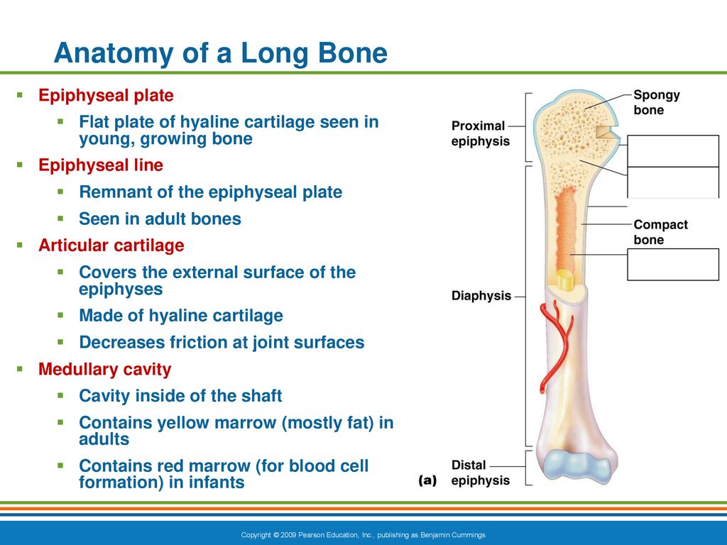
Bone and cartilage are two types of connective tissues. These joints are immovable (synarthrosis). Hyaline cartilage found at the end of long bones epiphyseal line long growth of bone occurs here during development where it is known as the growth plate or epiphyseal plate. Give examples for hyaline cartilage/ name the sites where it is present. A long bone has a shaft and 2 ends.
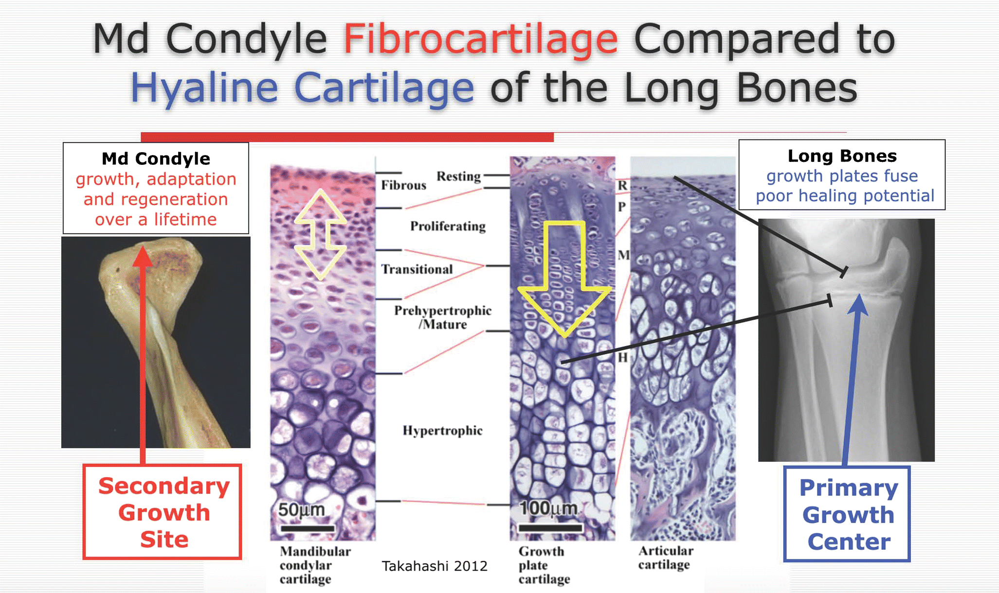
Figure 3 Part I Development And Physiology Of The Temporomandibular Joint Springerlink from media.springernature.com Parts of a long bone diagram quizlet / three types of cartilage are recognized based on differences in fiber composition:. Both bones and cartilages provide support and surfaces for the endochondral ossification produces the long bones such as humerus, radius, femur, and tibia by replacing the hyaline cartilage. It is the very hard substance that lines the bones in a joint; When a human finishes growing these parts fuse together. The epiphyseal plate is important because it is the site of bone growth. It gives the structures a definite but pliable form. The first indication of this process is the hypertrophy of the hyaline cartilage cells in the midshaft of the cartilagenous long bone. Connective tissue is subject to serious degenerative changes in joints because it has no blood supply.
The medullary cavity has a delicate membranous lining called the endosteum.
What structure in the diagram is the only place on a long bone not covered by the. Long bones, especially the femur and tibia, are subjected to most of the load during daily activities and they are crucial for skeletal mobility. The epiphyseal line is a remnant of an area that contained hyaline cartilage that grew during childhood to lengthen the bone. Labeled diagram of long bone. Hyaline cartilage is the most widespread and is the type that makes up the embryonic skeleton. Bone and cartilage are two types of connective tissues. On the epiphyseal side of the epiphyseal plate, cartilage is formed. Give examples for hyaline cartilage/ name the sites where it is present. The epiphyseal plate, a layer of hyaline cartilage, is replaced by osseous tissue as the organ grows in length. It can withstand compression forces, and yet it can bend. Long bone diagram hyaline cartilage. Both bones and cartilages provide support and surfaces for the endochondral ossification produces the long bones such as humerus, radius, femur, and tibia by replacing the hyaline cartilage. A long bone has two main regions:
How bones grow in length. End of the bone located farthest away from the midline 8. The epiphyseal plate, a layer of hyaline cartilage, is replaced by osseous tissue as the organ grows in length. The epiphyses, which are wider sections at each end of a long bone, are filled with spongy bone and red marrow. Hyaline cartilage is the most prevalent.
The Skeletal System Anatomy Of Long Bones Ppt Download from slideplayer.com The walls of the diaphysis are compact bone. Hyaline cartilage is the vital substance lining the bones in your joints. An example of a synchondrosis is the joint between the diaphysis and. Long bone diagram hyaline cartilage : Cartilage occurs where flexibility is required, while bone resists deformation. Labeled diagram of long bone. These joints are immovable (synarthrosis). | (a) ankle joint of the control group showing the.
How bones grow in length.
Hyaline cartilage is the most widespread and is the type that makes up the embryonic skeleton. Parts of a long bone diagram quizlet / three types of cartilage are recognized based on differences in fiber composition:. Long bones of the leg include the femur, tibia, fibula, metatarsals, and phalanges. Long bone diagram hyaline cartilage : The thigh bone (femur) is a long bone. The epiphyses, which are wider sections at each end of a long bone, are filled with spongy bone and red marrow. | (a) ankle joint of the control group showing the.the hyaline cartilage makes up most of the skeleton in the fetus because the skeleton is first laid down as hyaline cartilage. Hyaline cartilage is the most prevalent. The epiphyseal plate is a plate of hyaline cartilage found in bones that are growing in length. Long bone diagram hyaline cartilage : Once a child is born, his bones must grow longer and wider for him to grow bigger and taller. Hyaline cartilage exists on the sternal ends of the ribs, in the larynx, trachea, and bronchi, and on the articulating surfaces of bones. The presence of collagen fibres makes such structures and joints strong, but with limited mobility and flexibility.
When a human finishes growing these parts fuse together. The other two types are elastic cartilage and fibrocartilage. The material that takes the strain in the working parts. The epiphyses, which are wider sections at each end of a long bone, are filled with spongy bone and red marrow. The medullary cavity has a delicate membranous lining called the endosteum.
Anatomy Gross Anatomy Physiology Cells Cytology Cell Physiology Organelles Tissues Histology Organs Regional Anatomy Organ from www.apsubiology.org Hyaline cartilage exists on the sternal ends of the ribs, in the larynx, trachea, and bronchi, and on the articulating surfaces of bones. The diaphysis and the epiphysis. The epiphyseal plate is the area of growth in a long bone. End of a long bone. It is a layer of hyaline cartilage where ossification occurs in immature bones. Long bones of the leg include the femur, tibia, fibula, metatarsals, and phalanges. What structure in the diagram is the only place on a long bone not covered by the. Blood supply of long bones.
Long bone diagram hyaline cartilage :
When a human finishes growing these parts fuse together. How bones grow in length. Long bone diagram hyaline cartilage : Hundreds of these aggrecans are bound noncovalently by link proteins to long. That is, the whole bone is alive. The medullary cavity has a delicate membranous lining called the endosteum. The diaphysis and the epiphysis ( figure 6.3.1). The epiphyseal plate is a plate of hyaline cartilage found in bones that are growing in length. The diaphysis is the hollow, tubular shaft that runs between the proximal and distal ends of the bone. End of a long bone. End of the bone located farthest away from the midline 8. The first indication of this process is the hypertrophy of the hyaline cartilage cells in the midshaft of the cartilagenous long bone. Hyaline cartilage actually it is articular cartilage that lines the end of long bones.
| (a) ankle joint of the control group showing thethe hyaline cartilage makes up most of the skeleton in the fetus because the skeleton is first laid down as hyaline cartilage long bone diagram. | (a) ankle joint of the control group showing the.